| Protein: | gi|123997931, gi... |
| Organism: | synthetic construct |
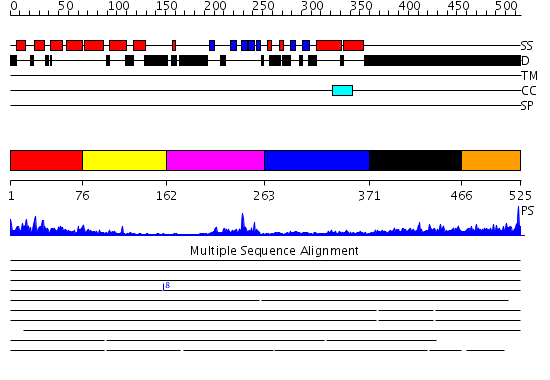
| Length: | 525 amino acids |
| Reference: | Drew K, et al. (2011) The proteome folding project: Proteome-scale prediction of structure and function. Genome Res. 2011 Sep 16 |
Listed below are up to the top 10 sequence alignment matches, by species, for the PSI-BLAST search against the protein sequence for gi|123997931, gi....
| Description | E-value | Query Range |
Subject Range |
|
|
0.0 | [1..525] | [1..525] |
|
|
0.0 | [1..525] | [1..525] |
|
|
1.0E-95 | [1..513] | [1..512] |
|
|
2.0E-94 | [1..525] | [1..523] |
|
|
6.0E-94 | [1..525] | [1..523] |
|
|
3.0E-92 | [14..525] | [148..659] |
|
|
2.0E-78 | [1..439] | [1..437] |
|
Region A: Residues: [1-75] |
      1          11         21         31         41         51
      |          |          |          |          |          |
    1 MPLFTANPFE QDVEKATNEY NTTEDWSLIM DICDKVGSTP NGAKDCLKAI MKRVNHKVPH  60
   61 VALQALTLLG ACVAN
|
| Detection Method: | |
| Confidence: | 40.69897 |
| Match: | 1x5bA |
| Description: | The solution structure of the VHS domain of human Signal transducing adaptor molecule 2 |
Matching Structure (courtesy of the PDB): |
|
|
Region A: Residues: [76-161] |
      1          11         21         31         41         51
      |          |          |          |          |          |
    1 CGKIFHLEVC SRDFATEVRA VIKNKAHPKV CEKLKSLMVE WSEEFQKDPQ FSLISATIKS  60
   61 MKEEGITFPP AGSQTVSAAA KNGTSS
|
| Detection Method: | |
| Confidence: | 3.67 |
| Match: | 1x5bA |
| Description: | The solution structure of the VHS domain of human Signal transducing adaptor molecule 2 |
| Matching Structure (courtesy of the PDB): |
|
|
Region A: Residues: [162-262] |
      1          11         21         31         41         51
      |          |          |          |          |          |
    1 NKNKEDEDIA KAIELSLQEQ KQQHTETKSL YPSSEIQLNN KVARKVRALY DFEAVEDNEL  60
   61 TFKHGEIIIV LDDSDANWWK GENHRGIGLF PSNFVTTNLN I
|
| Detection Method: | |
| Confidence: | 12.09691 |
| Match: | 1x2qA |
| Description: | Solution structure of the SH3 domain of the Signal transducing adaptor molecule 2 |
Matching Structure (courtesy of the PDB): |
|
|
Region A: Residues: [263-370] |
      1          11         21         31         41         51
      |          |          |          |          |          |
    1 ETEAAAVDKL NVIDDDVEEI KKSEPEPVYI DEDKMDRALQ VLQSIDPTDS KPDSQDLLDL  60
   61 EDICQQMGPM IDEKLEEIDR KHSELSELNV KVLEALELYN KLVNEAPV
|
| Detection Method: | |
| Confidence: | 10.522879 |
| Match: | 1bljA |
| Description: | NMR ENSEMBLE OF BLK SH2 DOMAIN, 20 STRUCTURES |
Matching Structure (courtesy of the PDB): |
|
| Term | Confidence | Notes |
| binding | 3.12724619324809 | bayes_pls_golite062009 |
| protein binding | 2.14586566310919 | bayes_pls_golite062009 |
| molecular adaptor activity | 1.33740973673697 | bayes_pls_golite062009 |
| SH3/SH2 adaptor activity | 1.2323381312171 | bayes_pls_golite062009 |
| ubiquitin-protein ligase activity | 1.02097165614658 | bayes_pls_golite062009 |
| signal transducer activity | 0.999269271132931 | bayes_pls_golite062009 |
| molecular transducer activity | 0.999269271132931 | bayes_pls_golite062009 |
| small conjugating protein ligase activity | 0.938262449967509 | bayes_pls_golite062009 |
| protein binding, bridging | 0.83823250804321 | bayes_pls_golite062009 |
| transcription regulator activity | 0.540818657754841 | bayes_pls_golite062009 |
| hydrolase activity | 0.539213162060111 | bayes_pls_golite062009 |
| nucleic acid binding | 0.476094076158571 | bayes_pls_golite062009 |
| DNA binding | 0.281643350069021 | bayes_pls_golite062009 |
| phosphoprotein binding | 0.16798254471727 | bayes_pls_golite062009 |
| acid-amino acid ligase activity | 0.125965304296204 | bayes_pls_golite062009 |
|
Region A: Residues: [371-465] |
      1          11         21         31         41         51
      |          |          |          |          |          |
    1 YSVYSKLHPP AHYPPASSGV PMQTYPVQSH GGNYMGQSIH QVTVAQSYSL GPDQIGPLRS  60
   61 LPPNVNSSVT AQPAQTSYLS TGQDTVSNPT YMNQN
|
| Detection Method: | |
| Confidence: | 1.06 |
| Match: | 1pk8A |
| Description: | Crystal Structure of Rat Synapsin I C Domain Complexed to Ca.ATP |
Matching Structure (courtesy of the PDB): |
|
|
Region A: Residues: [466-525] |
      1          11         21         31         41         51
      |          |          |          |          |          |
    1 SNLQSATGTT AYTQQMGMSV DMSSYQNTTS NLPQLAGFPV TVPAHPVAQQ HTNYHQQPLL  60
   61
|
| Detection Method: |
Shown below is our most confident prediction for this domain.
Click here to view all matches.
Found no confident structure predictions for this domain.