| Protein: | h0004690 |
| Organism: | SPECIES UNKNOWN |
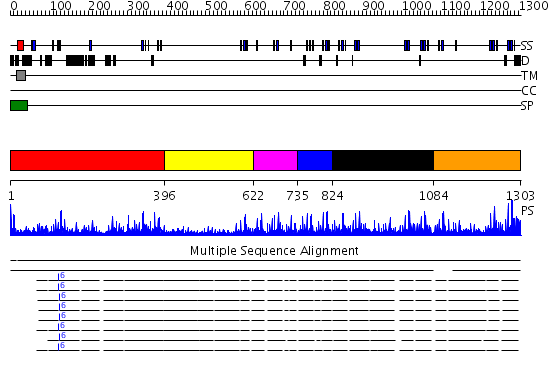
| Length: | 1303 amino acids |
| Reference: | Drew K, et al. (2011) The proteome folding project: Proteome-scale prediction of structure and function. Genome Res. 2011 Sep 16 |
Listed below are up to the top 10 sequence alignment matches, by species, for the PSI-BLAST search against the protein sequence for h0004690.
| Description | E-value | Query Range |
Subject Range |
|
|
0.0 | [1..1303] | [81..1382] |
|
|
0.0 | [68..1300] | [21..1187] |
|
|
0.0 | [70..1300] | [16..1182] |
|
|
0.0 | [96..1300] | [18..1158] |
|
Region A: Residues: [1-395] |
      1          11         21         31         41         51
      |          |          |          |          |          |
    1 MPGPRGAAGG LAPEMRGAGA AGLLALLLLL LLLLLGLGGR VEGGPAGERG AGGGGALARE  60
   61 RFKVVFAPVI CKRTCLKGQC RDSCQQGSNM TLIGENGHST DTLTGSGFRV VVCPLPCMNG 120
  121 GQCSSRNQCL CPPDFTGRFC QVPAGGAGGG TGGSGPGLSR TGALSTGALP PLAPEGDSVA 180
  181 SKHAIYAVQV IADPPGPGEG PPAQHAAFLV PLGPGQISAE VQAPPPVVNV RVHHPPEASV 240
  241 QVHRIESSNA ESAAPSQHLL PHPKPSHPRP PTQKPLGRCF QDTLPKQPCG SNPLPGLTKQ 300
  301 EDCCGSIGTA WGQSKCHKCP QLQYTGVQKP GPVRGEVGAD CPQGYKRLNS THCQDINECA 360
  361 MPGVCRHGDC LNNPGSYRCV CPPGHSLGPS RTQCI
|
| Detection Method: | |
| Confidence: | 15.69897 |
| Match: | 1n7dA |
| Description: | Low density lipoprotein (LDL) receptor YWTD domain; Low density lipoprotein (LDL) receptor, different EGF domains; Ligand-binding domain of low-density lipoprotein receptor |
Matching Structure (courtesy of the PDB): |
|
| Term | Confidence | Notes |
| receptor binding | 4.60755835611133 | bayes_pls_golite062009 |
| growth factor activity | 4.38305031783896 | bayes_pls_golite062009 |
| extracellular matrix structural constituent | 3.98005075037205 | bayes_pls_golite062009 |
| epidermal growth factor receptor binding | 2.63546895705146 | bayes_pls_golite062009 |
| ErbB-2 class receptor binding | 2.42443754640644 | bayes_pls_golite062009 |
| binding | 2.33940742543305 | bayes_pls_golite062009 |
| protein binding | 1.97546801934296 | bayes_pls_golite062009 |
| pattern binding | 1.68711415550213 | bayes_pls_golite062009 |
| glycosaminoglycan binding | 1.59362625477231 | bayes_pls_golite062009 |
| heparin binding | 1.18377533277052 | bayes_pls_golite062009 |
| ErbB-3 class receptor binding | 1.17748426017837 | bayes_pls_golite062009 |
| polysaccharide binding | 0.907482471490999 | bayes_pls_golite062009 |
| transcription regulator activity | 0.282532662545359 | bayes_pls_golite062009 |
| transcription factor binding | 0.25804698792789 | bayes_pls_golite062009 |
| transcription cofactor activity | 0.154111600670615 | bayes_pls_golite062009 |
|
Region A: Residues: [396-621] |
      1          11         21         31         41         51
      |          |          |          |          |          |
    1 ADKPEEKSLC FRLVSPEHQC QHPLTTRLTR QLCCCSVGKA WGARCQRCPT DGTAAFKEIC  60
   61 PAGKGYHILT SHQTLTIQGE SDFSLFLHPD GPPKPQQLPE SPSQAPPPED TEEERGVTTD 120
  121 SPVSEERSVQ QSHPTATTTP ARPYPELISR PSPPTMRWFL PDLPPSRSAV EIAPTQVTET 180
  181 DECRLNQNIC GHGECVPGPP DYSCHCNPGY RSHPQHRYCV DVNECE
|
| Detection Method: | |
| Confidence: | 3.08 |
| Match: | 1z1yA |
| Description: | Crystal structure of Methylated Pvs25, an ookinete protein from Plasmodium vivax |
Matching Structure (courtesy of the PDB): |
|
|
Region A: Residues: [622-734] |
      1          11         21         31         41         51
      |          |          |          |          |          |
    1 AEPCGPGRGI CMNTGGSYNC HCNRGYRLHV GAGGRSCVDL NECAKPHLCG DGGFCINFPG  60
   61 HYKCNCYPGY RLKASRPPVC EDIDECRDPS SCPDGKCENK PGSFKCIACQ PGY
|
| Detection Method: | |
| Confidence: | 5.0 |
| Match: | 1lmjA |
| Description: | Fibrillin-1 |
Matching Structure (courtesy of the PDB): |
|
| Term | Confidence | Notes |
| receptor binding | 4.60755835611133 | bayes_pls_golite062009 |
| growth factor activity | 4.38305031783896 | bayes_pls_golite062009 |
| extracellular matrix structural constituent | 3.98005075037205 | bayes_pls_golite062009 |
| epidermal growth factor receptor binding | 2.63546895705146 | bayes_pls_golite062009 |
| ErbB-2 class receptor binding | 2.42443754640644 | bayes_pls_golite062009 |
| binding | 2.33940742543305 | bayes_pls_golite062009 |
| protein binding | 1.97546801934296 | bayes_pls_golite062009 |
| pattern binding | 1.68711415550213 | bayes_pls_golite062009 |
| glycosaminoglycan binding | 1.59362625477231 | bayes_pls_golite062009 |
| heparin binding | 1.18377533277052 | bayes_pls_golite062009 |
| ErbB-3 class receptor binding | 1.17748426017837 | bayes_pls_golite062009 |
| polysaccharide binding | 0.907482471490999 | bayes_pls_golite062009 |
| transcription regulator activity | 0.282532662545359 | bayes_pls_golite062009 |
| transcription factor binding | 0.25804698792789 | bayes_pls_golite062009 |
| transcription cofactor activity | 0.154111600670615 | bayes_pls_golite062009 |
|
Region A: Residues: [735-823] |
      1          11         21         31         41         51
      |          |          |          |          |          |
    1 RSQGGGACRD VNECAEGSPC SPGWCENLPG SFRCTCAQGY APAPDGRSCL DVDECEAGDV  60
   61 CDNGICSNTP GSFQCQCLSG YHLSRDRSH
|
| Detection Method: | |
| Confidence: | 3.69897 |
| Match: | 1emnA |
| Description: | NMR STUDY OF A PAIR OF FIBRILLIN CA2+ BINDING EPIDERMAL GROWTH FACTOR-LIKE DOMAINS, MINIMIZED AVERAGE STRUCTURE |
Matching Structure (courtesy of the PDB): |
|
|
Region A: Residues: [824-1083] |
      1          11         21         31         41         51
      |          |          |          |          |          |
    1 CEDIDECDFP AACIGGDCIN TNGSYRCLCP QGHRLVGGRK CQDIDECSQD PSLCLPHGAC  60
   61 KNLQGSYVCV CDEGFTPTQD QHGCEEVEQP HHKKECYLNF DDTVFCDSVL ATNVTQQECC 120
  121 CSLGAGWGDH CEIYPCPVYS SAEFHSLCPD GKGYTQDNNI VNYGIPAHRD IDECMLFGSE 180
  181 ICKEGKCVNT QPGYECYCKQ GFYYDGNLLE CVDVDECLDE SNCRNGVCEN TRGGYRCACT 240
  241 PPAEYSPAQR QCLSPEEMDV
|
| Detection Method: | |
| Confidence: | 4.29 |
| Match: | 1z1yA |
| Description: | Crystal structure of Methylated Pvs25, an ookinete protein from Plasmodium vivax |
| Matching Structure (courtesy of the PDB): |
|
|
Region A: Residues: [1084-1303] |
      1          11         21         31         41         51
      |          |          |          |          |          |
    1 DECQDPAACR PGRCVNLPGS YRCECRPPWV PGPSGRDCQL PESPAERAPE RRDVCWSQRG  60
   61 EDGMCAGPLA GPALTFDDCC CRQGRGWGAQ CRPCPPRGAG SHCPTSQSES NSFWDTSPLL 120
  121 LGKPPRDEDS SEEDSDECRC VSGRCVPRPG GAVCECPGGF QLDASRARCV DIDECRELNQ 180
  181 RGLLCKSERC VNTSGSFRCV CKAGFARSRP HGACVPQRRR
|
| Detection Method: | |
| Confidence: | 5.522879 |
| Match: | 1uzjA |
| Description: | Integrin binding cbEGF22-TB4-cbEGF33 fragment of human fibrillin-1, holo form. |
Matching Structure (courtesy of the PDB): |
|