| Protein: | mat-3 |
| Organism: | Caenorhabditis elegans |
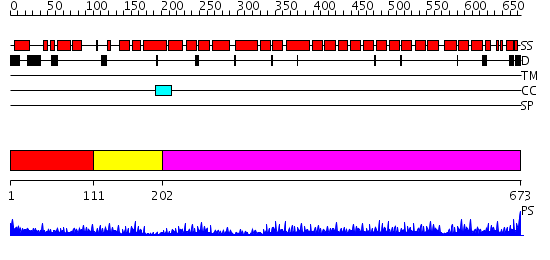
| Length: | 673 amino acids |
| Reference: | Drew K, et al. (2011) The proteome folding project: Proteome-scale prediction of structure and function. Genome Res. 2011 Sep 16 |
Listed below are up to the top 10 sequence alignment matches, by species, for the PSI-BLAST search against the protein sequence for mat-3.
| Description | E-value | Query Range |
Subject Range |
|
|
417.0 | [0..57] | [672..25] |
|
Region A: Residues: [1-110] |
      1          11         21         31         41         51
      |          |          |          |          |          |
    1 MNVSFSTPVQ NSTASRLALI QQAAARSGAL PPAGQYRGSK LSPFDVSQMS GVSISMVERA  60
   61 KINGPEFVEE LEWLRQQTTS RCFLDAEMWT NEILAHLPDK WCAPNTLNLY
|
| Detection Method: |
Shown below is our most confident de novo (Rosetta) prediction for this domain.
Click here to view all matches.
Found no confident structure predictions for this domain.
|
Region A: Residues: [111-201] |
      1          11         21         31         41         51
      |          |          |          |          |          |
    1 NQVSELVLDN TRSPMASPAS NSLYAPGEDQ MPTVKRNHTS RFAQSLIKNK EFRRAAFFLE  60
   61 KTMNGNKLDH FLHFRCLFLA YYQEHLENDA E
|
| Detection Method: | |
| Confidence: | 3.154902 |
| Match: | 1zbpA |
| Description: | Crystal Structure of the Hypothetical protein VPA1032 from Vibrio parahaemolyticus, Northeast Structural Genomics Target VpR44 |
Matching Structure (courtesy of the PDB): |
|
|
Region A: Residues: [202-673] |
      1          11         21         31         41         51
      |          |          |          |          |          |
    1 GIERKTSFAE ERSPFSLLYQ RMEDKKLREN EDVWFEYLMG LLEVELGLKD LAEKSFRNVV  60
   61 IREPRIWPAW EALSRLIADI EDADKFVTSA EVKSLWMGDW FMTLVLQRFH QHSMAIQKAE 120
  121 QLVTRGMTGL PMIITKIAAC SNARHDHDQA ISNFEDVRKA DPYRLGDLHL LSDSLYIRND 180
  181 QKKLSTLAIE VYKVHKFRWE TCCIVANYHA IRRDSEHAIK FFQRALRLNP GLAALWVLIG 240
  241 HEFMEMKNNA AACVSYRRAI EIDPADHRGW YGLGQMYDIM KMPAYALFYY QEAQKCKPHD 300
  301 SRLLVALGDI YSKLNRIEDA EKCFTGAYLF GDVEGNALWS LAKLHERYSD DNKAAQAFEV 360
  361 FLVVYELVTS AEEKIIYAIA FLANHFFKIE DFDKASEYAT KCLAFETLCQ EGNRLFREIA 420
  421 KIQARESRLP VEEAPGPSNA SAAGGQEAMD TEEAPQEGGE EEMSEGEDDF SF
|
| Detection Method: | |
| Confidence: | 59.69897 |
| Match: | 1w3bA |
| Description: | The superhelical TPR domain of O-linked GlcNAc transferase reveals structural similarities to importin alpha. |
Matching Structure (courtesy of the PDB): |
|
| Term | Confidence | Notes |
| ubiquitin-protein ligase activity | 6.63746543212322 | bayes_pls_golite062009 |
| small conjugating protein ligase activity | 6.37753885031863 | bayes_pls_golite062009 |
| acid-amino acid ligase activity | 5.68162115570289 | bayes_pls_golite062009 |
| ligase activity, forming carbon-nitrogen bonds | 4.89523122092864 | bayes_pls_golite062009 |
| binding | 2.57186536498101 | bayes_pls_golite062009 |
| ligase activity | 1.72049482453565 | bayes_pls_golite062009 |
| protein binding | 1.65083457326273 | bayes_pls_golite062009 |
| hydrolase activity | 0.619711232527193 | bayes_pls_golite062009 |
| nucleic acid binding | 0.41706917101675 | bayes_pls_golite062009 |
| transcription regulator activity | 0.194719472400022 | bayes_pls_golite062009 |
| DNA binding | 0.17736976899009 | bayes_pls_golite062009 |