| Protein: | pdfr-1 |
| Organism: | Caenorhabditis elegans |
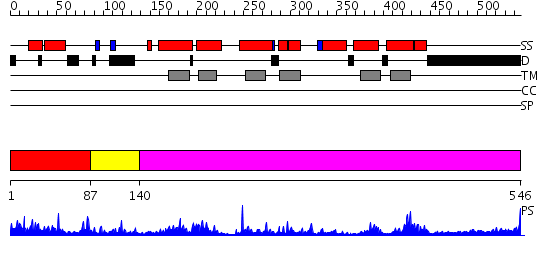
| Length: | 546 amino acids |
| Reference: | Drew K, et al. (2011) The proteome folding project: Proteome-scale prediction of structure and function. Genome Res. 2011 Sep 16 |
|
Region A: Residues: [1-86] |
      1          11         21         31         41         51
      |          |          |          |          |          |
    1 MADATSPFNV SILDNSTKLS EMVESGWNVL ASTSVQAFNE AMDVLEESYP LCKKMLDHNN  60
   61 LFPERDPNDT RIWCNATYDT VLCWPP
|
| Detection Method: | |
| Confidence: | 19.522879 |
| Match: | 1ijyA |
| Description: | Frizzled 8 (FZ8) |
Matching Structure (courtesy of the PDB): |
|
| Term | Confidence | Notes |
| transmembrane receptor activity | 4.38708793497137 | bayes_pls_golite062009 |
| receptor activity | 4.1046853496316 | bayes_pls_golite062009 |
| signal transducer activity | 3.17034085692897 | bayes_pls_golite062009 |
| molecular transducer activity | 3.17034085692897 | bayes_pls_golite062009 |
| Wnt receptor activity | 2.95244120669902 | bayes_pls_golite062009 |
| binding | 2.18217693324765 | bayes_pls_golite062009 |
| protein binding | 2.15165760199286 | bayes_pls_golite062009 |
| Wnt-protein binding | 2.04664201899031 | bayes_pls_golite062009 |
|
Region A: Residues: [87-139] |
      1          11         21         31         41         51
      |          |          |          |          |          |
    1 TPANSSVTLQ CPHMKGLDPN KNITKDCHVS GVWSGRNAGE MGPTLPGWTN FTM
|
| Detection Method: | |
| Confidence: | 1.84 |
| Match: | 1u34A |
| Description: | 3D NMR structure of the first extracellular domain of CRFR-2beta, a type B1 G-protein coupled receptor |
Matching Structure (courtesy of the PDB): |
|
|
Region A: Residues: [140-546] |
      1          11         21         31         41         51
      |          |          |          |          |          |
    1 CYTDEVIYIM QNLNNESLTI AQEVARNARK LEFVGLGLSL VSLILAISIF SYFRRLRVFR  60
   61 NLLHLHLMIA MLMVVILRLV LYIDLIFTGE NGPHTNSAEG KTINTMPIVC EGMFFFLEYF 120
  121 KTVTFCWMFL EGIYLNNQIV FGFFNSEPKL LPYFIAGYGI PLVHTMLWLL VVLIKKDFKV 180
  181 ERCLGSYYLE PEFWILDGPR MAELVINLFF ICNVIRVLYS KVRESNNTSE AGLKKSVKAA 240
  241 MMLLPLLGVP NIMQTIPFAP TRDNIMVFAV WTYTASFTYM YQGLMVASIY CFTNKEVNHV 300
  301 LKTFYARYRL LHKSQNELRR GSRSVASHYA AKNGTANASA PQTNNADEFG KLSPFPSRSK 360
  361 KGSDDSTTKL MKDAVMEEEK NANNNGYGSA GEMTPLREGS NRSTKSP
|
| Detection Method: | |
| Confidence: | 49.69897 |
| Match: | 1f88A |
| Description: | Rhodopsin |
Matching Structure (courtesy of the PDB): |
|