| Protein: | PH4alphaPV-PA |
| Organism: | Drosophila melanogaster |
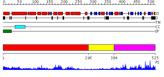
| Length: | 525 amino acids |
| Reference: | Drew K, et al. (2011) The proteome folding project: Proteome-scale prediction of structure and function. Genome Res. 2011 Sep 16 |
Listed below are up to the top 10 sequence alignment matches, by species, for the PSI-BLAST search against the protein sequence for PH4alphaPV-PA.
| Description | E-value | Query Range |
Subject Range |
|
|
404.0 | [0..8] | [525..1] |
|
Region A: Residues: [1-295] |
      1          11         21         31         41         51
      |          |          |          |          |          |
    1 MRSSKWLLPV RVLLTYQVLL LLLRQAKGEE SHCTSVAGMV KLLDLEAQLI DNLEDYAVEL  60
   61 EKKLQTVRRS ITSLRLENDK ARSSTEEYLS NPLNSFSLIR RMNRDWIIWQ LYMDDPVGIS 120
  121 QVERIQELRE HMPTHTDVEE AVTALDRIQS TYGLKVPEIS HGFLNGKQYN VSLTVLDTYA 180
  181 MGQILFDQKN YLAAASWIYQ SVVLMEAFSM AAPLEISKNE VRMVYAETLL KLNQHADALK 240
  241 VVNIALTDNP HDIKLLLKKS EIETMIRTGT NNAPPVKVQA TGVPTAYQIG CRGQF
|
| Detection Method: | |
| Confidence: | 30.522879 |
| Match: | 1w3bA |
| Description: | The superhelical TPR domain of O-linked GlcNAc transferase reveals structural similarities to importin alpha. |
Matching Structure (courtesy of the PDB): |
|
| Term | Confidence | Notes |
| procollagen-proline 4-dioxygenase activity | 7.0422525853653 | bayes_pls_golite062009 |
| procollagen-proline dioxygenase activity | 7.0422525853653 | bayes_pls_golite062009 |
| peptidyl-proline dioxygenase activity | 7.0422525853653 | bayes_pls_golite062009 |
| peptidyl-proline 4-dioxygenase activity | 7.0422525853653 | bayes_pls_golite062009 |
| oxidoreductase activity, acting on paired donors, with incorporation or reduction of molecular oxygen, 2-oxoglutarate as one donor, and incorporation of one atom each of oxygen into both donors | 5.01373106146099 | bayes_pls_golite062009 |
| binding | 2.09193012129784 | bayes_pls_golite062009 |
| protein binding | 1.52807685989528 | bayes_pls_golite062009 |
| oxidoreductase activity, acting on NADH or NADPH, with oxygen as acceptor | 0.534990536504941 | bayes_pls_golite062009 |
|
Region A: Residues: [296-383] |
      1          11         21         31         41         51
      |          |          |          |          |          |
    1 PPSADSKLYC LYNRTTSPFL ILAPLKMELV GLDPYMVLYH DVLSPKEIKE LQGMATPGLK  60
   61 RATVYQASSG RNEVVKTRTS KVAWFPDG
|
| Detection Method: | |
| Confidence: | 5.39794 |
| Match: | 2c2lA |
| Description: | Crystal structure of the CHIP U-box E3 ubiquitin ligase |
Matching Structure (courtesy of the PDB): |
|
|
Region A: Residues: [384-525] |
      1          11         21         31         41         51
      |          |          |          |          |          |
    1 YNPLTVRLNA RISDMTGFNL YGSEMLQLMN YGLGGHYDQH YDFFNKTNSN MTAMSGDRIA  60
   61 TVLFYLTDVE QGGATVFPNI RKAVFPQRGS VVMWYNLKDN GQIDTQTLHA ACPVIVGSKW 120
  121 VCNKWIRERE QIFSRPCLKK RM
|
| Detection Method: | |
| Confidence: | 4.34 |
| Match: | 2hbtA |
| Description: | No description for 2hbtA was found. |