| Protein: | SERA2_ARATH |
| Organism: | Arabidopsis thaliana |
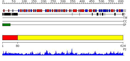
| Length: | 624 amino acids |
| Reference: | Drew K, et al. (2011) The proteome folding project: Proteome-scale prediction of structure and function. Genome Res. 2011 Sep 16 |
Listed below are up to the top 10 sequence alignment matches, by species, for the PSI-BLAST search against the protein sequence for SERA2_ARATH.
| Description | E-value | Query Range |
Subject Range |
|
|
503.0 | [0..84] | [624..3] |
|
Region A: Residues: [1-79] |
      1          11         21         31         41         51
      |          |          |          |          |          |
    1 MAFSSSCSSV KAVNSRWTSP SPSPSSRFAV LPAFLHRRYA TSVKLTAISA ALKTVEQTTL  60
   61 TEDNRFSTVG SDSDEYNPT
|
| Detection Method: | |
| Confidence: | 82.69897 |
| Match: | 1b3rA |
| Description: | S-adenosylhomocystein hydrolase |
Matching Structure (courtesy of the PDB): |
|
|
Region A: Residues: [80-624] |
      1          11         21         31         41         51
      |          |          |          |          |          |
    1 LPKPRILVTE KLGEAGVNLL REFGDVDCSY DLSPEDLKKK VAESDALIVR SGTKVTREVF  60
   61 EAAKGRLKVV GRAGVGIDNV DLQAATEHGC LVVNAPTANT VAAAEHGIAL LASMARNVAQ 120
  121 ADASIKAGKW ERSKYVGVSL VGKTLAVMGF GKVGTEVARR AKGLGMTVIS HDPYAPADRA 180
  181 RALGVDLVSF DQAISTADFV SLHMPLTPAT KKVFNDETFS KMKKGVRLIN VARGGVIDED 240
  241 ALVRALDAGI VAQAALDVFC EEPPSKDSRL IQHENVTVTP HLGASTKEAQ EGVAIEIAEA 300
  301 VAGALKGELS ATAVNAPMVA PEVLSELTPY IVLAEKLGRL AVQLASGGKG VQSIRVVYRS 360
  361 ARDRDDLDTR LLRAMITKGI IEPISDSYVN LVNADFIAKQ KGLRISEERM VVDSSPEYPV 420
  421 DSIQVQILNV ESNFAGAVSD AGDISIEGKV KYGVPHLTCV GSFGVDVSLE GNLILCRQVD 480
  481 QPGMIGQVGN ILGEQNVNVN FMSVGRTVLR KQAIMAIGVD EEPDNKTLER IGGVSAIEEF 540
  541 VFLKL
|
| Detection Method: | |
| Confidence: | 110.0 |
| Match: | 1ygyA |
| Description: | Crystal Structure of D-3-Phosphoglycerate dehydrogenase From Mycobacterium tuberculosis |
Matching Structure (courtesy of the PDB): |
|