| Protein: | feh-1 |
| Organism: | Caenorhabditis elegans |
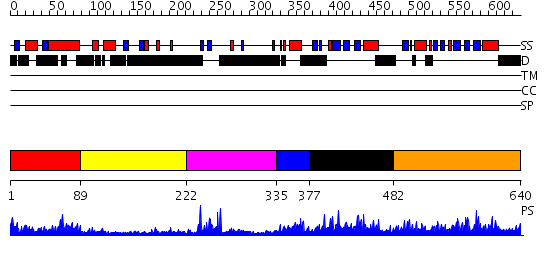
| Length: | 640 amino acids |
| Reference: | Drew K, et al. (2011) The proteome folding project: Proteome-scale prediction of structure and function. Genome Res. 2011 Sep 16 |
Listed below are up to the top 10 sequence alignment matches, by species, for the PSI-BLAST search against the protein sequence for feh-1.
| Description | E-value | Query Range |
Subject Range |
|
|
528.0 | [0..55] | [637..104] |
|
Region A: Residues: [1-88] |
      1          11         21         31         41         51
      |          |          |          |          |          |
    1 MREGTPRVRI EVNKGSNRPS QFVSESEEQR LQRVQSRDSD TATMNFVTDD RMMEEHDEYS  60
   61 AFKEAYEEEE SREYVIENGV KRLVNQHY
|
| Detection Method: |
Shown below is our most confident prediction for this domain.
Click here to view all matches.
Found no confident structure predictions for this domain.
|
Region A: Residues: [89-221] |
      1          11         21         31         41         51
      |          |          |          |          |          |
    1 DSRGYSSAGR GQKGRREEER RRNMAVDYSS QDFRQSLAAI DQASRDGAIS RGEDGVRVRQ  60
   61 IGDTTIMTEH RDPYSFYWQL DQARREAESP PRRPPPTDYV IDEEEEVLET VYSPEADDVM 120
  121 FENQYRRPVR HPL
|
| Detection Method: |
Shown below is our most confident de novo (Rosetta) prediction for this domain.
Click here to view all matches.
Found no confident structure predictions for this domain.
|
Region A: Residues: [222-334] |
      1          11         21         31         41         51
      |          |          |          |          |          |
    1 PPPPPIMEEE PKDLPPGWEK HEDPQGYSYY WHVDSGTIQR QPPPPVNRET QADAPPPQII  60
   61 QLPPQQPVIE EHAFKQTTTK RRIEQDEMSE REIEDVAMIE NGDTYHKPVR FAV
|
| Detection Method: | |
| Confidence: | 10.221849 |
| Match: | 2f21A |
| Description: | No description for 2f21A was found. |
|
Region A: Residues: [335-376] |
      1          11         21         31         41         51
      |          |          |          |          |          |
    1 RSLGWTDISE DELTAEKSSR AVNRAIVDLT TRSDIDSIPK WG
|
| Detection Method: | |
| Confidence: | 4.09691 |
| Match: | 2dk7A |
| Description: | No description for 2dk7A was found. |
|
Region A: Residues: [377-481] |
      1          11         21         31         41         51
      |          |          |          |          |          |
    1 DGRELIMELD DNELALLDPD SMNVIHSERI QAIRVWGVGR DNGRDFAYVS RDRGTRRFMC  60
   61 HVFRCDTSAK TIANTLRDIC KRLMLHRRPS SLHAIESGEK RIVRS
|
| Detection Method: | |
| Confidence: | 2.72 |
| Match: | 1m7eA |
| Description: | Crystal structure of the phosphotyrosine binding domain(PTB) of mouse Disabled 2(Dab2):implications for Reeling signaling |
Matching Structure (courtesy of the PDB): |
|
|
Region A: Residues: [482-640] |
      1          11         21         31         41         51
      |          |          |          |          |          |
    1 EGLTAPIDEP RKVIRCHFLG VTQVPKATGI EILNEAVDRL VSQVRSERWI LADVSIAPST  60
   61 IAIVEVNGQQ IAECRVRYLS FLGIGRDVKH CAFIMQTSSE SFMCYVFHVE PNAAAMAKMV 120
  121 EAACKLRYQK VLDAHSSSRH HSGMSIHGQH PPSTYHGMD
|
| Detection Method: | |
| Confidence: | 44.522879 |
| Match: | 1wguA |
| Description: | Solution Structure of the C-terminal Phosphotyrosine Interaction Domain of APBB2 from Mouse |
Matching Structure (courtesy of the PDB): |
|