| Protein: | gi|28573155, gi|... |
| Organism: | Drosophila melanogaster |
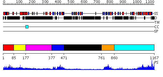
| Length: | 1167 amino acids |
| Reference: | Drew K, et al. (2011) The proteome folding project: Proteome-scale prediction of structure and function. Genome Res. 2011 Sep 16 |
Listed below are up to the top 10 sequence alignment matches, by species, for the PSI-BLAST search against the protein sequence for gi|28573155, gi|....
| Description | E-value | Query Range |
Subject Range |
|
|
484.0 | [0..194] | [1167..1] |
|
Region A: Residues: [1-84] |
      1          11         21         31         41         51
      |          |          |          |          |          |
    1 MTREEEEEVR SLLSRHQERI MLDLGSTDLL AVLVKNSVLS QSEEDLLLKP YNAVTPTPSA  60
   61 AASATVEPPG SAPSLTKRKL SAVA
|
| Detection Method: |
Shown below is our most confident de novo (Rosetta) prediction for this domain.
Click here to view all matches.
Found no confident structure predictions for this domain.
|
Region A: Residues: [85-176] |
      1          11         21         31         41         51
      |          |          |          |          |          |
    1 SSGSTASGGS AGSGGSGSSR CASVNGDANC DSRSLTPSGG GPCSGLNDAE ILRVQCSNLI  60
   61 DIIAKNGFEK FKQFCYAIEC ECPQLIEDLI ND
|
| Detection Method: | |
| Confidence: | 17.30103 |
| Match: | 1ujvA |
| Description: | Solution structure of the second PDZ domain of human membrane associated guanylate kinase inverted-2 (MAGI-2) |
Matching Structure (courtesy of the PDB): |
|
| Term | Confidence | Notes |
| binding | 2.6196222853442 | bayes_pls_golite062009 |
| protein binding | 2.35875654816667 | bayes_pls_golite062009 |
| signal transducer activity | 1.06018756723145 | bayes_pls_golite062009 |
| molecular transducer activity | 1.06018756723145 | bayes_pls_golite062009 |
| protein domain specific binding | 0.64214865642772 | bayes_pls_golite062009 |
| cell adhesion molecule binding | 0.403457741099606 | bayes_pls_golite062009 |
| receptor binding | 0.359972518335054 | bayes_pls_golite062009 |
| PDZ domain binding | 0.0377679976799299 | bayes_pls_golite062009 |
|
Region A: Residues: [177-376] |
      1          11         21         31         41         51
      |          |          |          |          |          |
    1 RLKTDAANLE DSRDGEKMQK EIETIDYIDK EKENDRNRRA VSYGGDATSS PHSATIPRAA  60
   61 PRRVALMKDP PPPPPPKPQL GSKADSVSHL ASKYPQSEYN LIQKIDSNST LTAPAQYQPQ 120
  121 NFYANTGTIS STNGYGSLLC HASSTTSPLA VRKRDKLLHR FSDAATLGRK LKKKKNTNRT 180
  181 CRSMTEAIEM LADPVIEDEF
|
| Detection Method: |
Shown below is our most confident de novo (Rosetta) prediction for this domain.
Click here to view all matches.
Found no confident structure predictions for this domain.
|
Region A: Residues: [377-470] |
      1          11         21         31         41         51
      |          |          |          |          |          |
    1 FGDRTTWEYH TVAVTRVPGY GFGIAVSGGR DNPHFANGDP SIAVSDVLKG GPAEDRLQVN  60
   61 DRIISVNGVS LENVEYATAV QVLRDSGNTV QLVV
|
| Detection Method: | |
| Confidence: | 23.39794 |
| Match: | 2h2bA |
| Description: | No description for 2h2bA was found. |
|
Region A: Residues: [471-760] |
      1          11         21         31         41         51
      |          |          |          |          |          |
    1 KRRVPLNPIN AAGAVQHQHS HSLSSVGLMA NGSGGVAPTP ITSLSQPNSL NSSLVQNASS  60
   61 GQPIKVTLTK GGKKDDYGVV LGCRLFVKEI SSKAREQLNA NGYSLQEGDI ITRIHNTNCG 120
  121 DTMSLKEAKK IIDGCKERLN LVVLRDITNQ TAVSQLNLNN SASHQASGNI YATHQPQVSG 180
  181 CSSSNNNLED PYLPGGASYS SQNLYVQPPT RTSNGPNING NGLNDEKSNL TPRGRSRGPI 240
  241 MDGVSLQQLD RPVTPTRGRS AAIDEPPRPP PPRGSSGGAA QEDFYSSRRQ
|
| Detection Method: | |
| Confidence: | 3.59 |
| Match: | 1z87A |
| Description: | solution structure of the split PH-PDZ Supramodule of alpha-Syntrophin |
Matching Structure (courtesy of the PDB): |
|
|
Region A: Residues: [761-859] |
      1          11         21         31         41         51
      |          |          |          |          |          |
    1 LYEERQSAEP RFISFQKEGS VGIRLTGGNE AGIFVTAVQP GSPASLQGLM PGDKILKVND  60
   61 MDMNGVTREE AVLFLLSLQD RIDLIVQYCK EEYDEVVTN
|
| Detection Method: | |
| Confidence: | 19.69897 |
| Match: | 1um7A |
| Description: | Solution structure of the third PDZ domain of synapse-associated protein 102 |
Matching Structure (courtesy of the PDB): |
|
| Term | Confidence | Notes |
| binding | 2.6196222853442 | bayes_pls_golite062009 |
| protein binding | 2.35875654816667 | bayes_pls_golite062009 |
| signal transducer activity | 1.06018756723145 | bayes_pls_golite062009 |
| molecular transducer activity | 1.06018756723145 | bayes_pls_golite062009 |
| protein domain specific binding | 0.64214865642772 | bayes_pls_golite062009 |
| cell adhesion molecule binding | 0.403457741099606 | bayes_pls_golite062009 |
| receptor binding | 0.359972518335054 | bayes_pls_golite062009 |
| PDZ domain binding | 0.0377679976799299 | bayes_pls_golite062009 |
|
Region A: Residues: [860-1167] |
      1          11         21         31         41         51
      |          |          |          |          |          |
    1 QRGDSFHIKT HFHCDNPSKG EMAFKAGDVF RVIDTLHNGV VGSWQVLKIG RGHQEMQRGV  60
   61 IPNKSRAEEL ATAQFNATKK EMNANESRGN FFRRRRSTHR RSKSLSRENW DDVVFSDSIS 120
  121 KFPAYERVVL RHPGFVRPVV LFGPVSDLAR ERLAKDFPDK FSTPLQDDDK SAATSGKCRI 180
  181 VRLSNIRDVM DRGKHALLDI TPNAVDRLNY AQFYPVVIFL KTDSKHVIKQ LRHGLPKAAH 240
  241 KSSKKLLEQC QKLERVWSHI FSTQIALSDE ESWYRKLRDS IDLQQSGAVW MSESKTKTKY 300
  301 ISSWSFPA
|
| Detection Method: | |
| Confidence: | 41.39794 |
| Match: | 1kjwA |
| Description: | Psd-95; Guanylate kinase-like domain of Psd-95 |
Matching Structure (courtesy of the PDB): |
|