| Protein: | aralar1-PC |
| Organism: | Drosophila melanogaster |
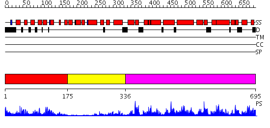
| Length: | 695 amino acids |
| Reference: | Drew K, et al. (2011) The proteome folding project: Proteome-scale prediction of structure and function. Genome Res. 2011 Sep 16 |
Listed below are up to the top 10 sequence alignment matches, by species, for the PSI-BLAST search against the protein sequence for aralar1-PC.
| Description | E-value | Query Range |
Subject Range |
|
|
483.0 | [0..15] | [695..2] |
|
Region A: Residues: [1-174] |
      1          11         21         31         41         51
      |          |          |          |          |          |
    1 MPMHIPFPFN WIPTLPVARC QESPSLLKRA GTEKLREVFL KYASIQKNGE HYMTSEDFVR  60
   61 KFLGLFSESA FNDESVRLLA NIADTSKDGL ISFSEFQAFE GLLCTPDALY RTAFQLFDRK 120
  121 GNGTVSYADF ADVVQKTELH SKIPFSLDGP FIKRYFGDKK QRLINYAEFT QLLH
|
| Detection Method: | |
| Confidence: | 41.522879 |
| Match: | 1zuzA |
| Description: | No description for 1zuzA was found. |
| Term | Confidence | Notes |
| calcium ion binding | 3.79609619542776 | bayes_pls_golite062009 |
| binding | 2.74623080162785 | bayes_pls_golite062009 |
| metal ion transmembrane transporter activity | 2.61874575467092 | bayes_pls_golite062009 |
| cytoskeletal protein binding | 2.59226289545114 | bayes_pls_golite062009 |
| ion transmembrane transporter activity | 2.57032504567157 | bayes_pls_golite062009 |
| cation transmembrane transporter activity | 2.55650624542181 | bayes_pls_golite062009 |
| substrate-specific transmembrane transporter activity | 2.37465721152046 | bayes_pls_golite062009 |
| structural molecule activity | 2.02132683356917 | bayes_pls_golite062009 |
| potassium channel activity | 1.89948827776974 | bayes_pls_golite062009 |
| motor activity | 1.81368053536929 | bayes_pls_golite062009 |
| voltage-gated potassium channel activity | 1.81135503818602 | bayes_pls_golite062009 |
| structural constituent of muscle | 1.6840107470242 | bayes_pls_golite062009 |
| actin binding | 1.50831300191117 | bayes_pls_golite062009 |
| cation channel activity | 1.49312394054212 | bayes_pls_golite062009 |
| signal transducer activity | 1.38521870126745 | bayes_pls_golite062009 |
| molecular transducer activity | 1.38521870126745 | bayes_pls_golite062009 |
| ion binding | 1.36376383271837 | bayes_pls_golite062009 |
| metal ion binding | 1.34827363853601 | bayes_pls_golite062009 |
| cation binding | 1.34827363853601 | bayes_pls_golite062009 |
| transcription regulator activity | 1.22886396511326 | bayes_pls_golite062009 |
| protein binding | 1.16246005294206 | bayes_pls_golite062009 |
| transporter activity | 1.15928534291452 | bayes_pls_golite062009 |
| microfilament motor activity | 1.0725773039399 | bayes_pls_golite062009 |
| channel activity | 1.05847414989139 | bayes_pls_golite062009 |
| passive transmembrane transporter activity | 1.05847414989139 | bayes_pls_golite062009 |
| ion channel activity | 1.0369591011376 | bayes_pls_golite062009 |
| substrate-specific channel activity | 1.03529144716694 | bayes_pls_golite062009 |
| voltage-gated cation channel activity | 0.986621437232479 | bayes_pls_golite062009 |
| transmembrane transporter activity | 0.945691505472547 | bayes_pls_golite062009 |
| voltage-gated ion channel activity | 0.910148857310729 | bayes_pls_golite062009 |
| voltage-gated channel activity | 0.896351900356978 | bayes_pls_golite062009 |
| gated channel activity | 0.855502499003241 | bayes_pls_golite062009 |
| tubulin binding | 0.851404549871251 | bayes_pls_golite062009 |
| nucleic acid binding | 0.849623084382492 | bayes_pls_golite062009 |
| actin-dependent ATPase activity | 0.83859958498483 | bayes_pls_golite062009 |
| DNA binding | 0.81633153359201 | bayes_pls_golite062009 |
| amine transmembrane transporter activity | 0.717054504236607 | bayes_pls_golite062009 |
| substrate-specific transporter activity | 0.693724160233023 | bayes_pls_golite062009 |
| microtubule binding | 0.482216984478791 | bayes_pls_golite062009 |
| myosin binding | 0.41761909113528 | bayes_pls_golite062009 |
| 0.34446792626966 | bayes_pls_golite062009 | |
| channel regulator activity | 0.32809521759917 | bayes_pls_golite062009 |
| protein complex binding | 0.278602833762221 | bayes_pls_golite062009 |
| active transmembrane transporter activity | 0.246383459363256 | bayes_pls_golite062009 |
| myosin heavy chain binding | 0.15875878144391 | bayes_pls_golite062009 |
| G-protein beta/gamma-subunit binding | 0.13723868253862 | bayes_pls_golite062009 |
| transcription factor binding | 0.02456682442567 | bayes_pls_golite062009 |
| actin filament binding | 0.0239092123185181 | bayes_pls_golite062009 |
|
Region A: Residues: [175-335] |
      1          11         21         31         41         51
      |          |          |          |          |          |
    1 DFHEEHAMEA FRSKDPAGTG FISPLDFQDI IVNVKRHLLT PGVRDNLVSV TEGHKVSFPY  60
   61 FIAFTSLLNN MELIKQVYLH ATEGSRTDMI TKDQILLAAQ TMSQITPLEI DILFHLAGAV 120
  121 HQAGRIDYSD LSNIAPEHYT KHMTHRLAEI KAVESPADRS A
|
| Detection Method: | |
| Confidence: | 4.36 |
| Match: | 2f2oA |
| Description: | Structure of calmodulin bound to a calcineurin peptide: a new way of making an old binding mode |
Matching Structure (courtesy of the PDB): |
|
|
Region A: Residues: [336-695] |
      1          11         21         31         41         51
      |          |          |          |          |          |
    1 FIQVLESSYR FTLGSFAGAV GATVVYPIDL VKTRMQNQRA GSYIGEVAYR NSWDCFKKVV  60
   61 RHEGFMGLYR GLLPQLMGVA PEKAIKLTVN DLVRDKLTDK KGNIPTWAEV LAGGCAGASQ 120
  121 VVFTNPLEIV KIRLQVAGEI ASGSKIRAWS VVRELGLFGL YKGARACLLR DVPFSAIYFP 180
  181 TYAHTKAMMA DKDGYNHPLT LLAAGAIAGV PAASLVTPAD VIKTRLQVVA RSGQTTYTGV 240
  241 WDATKKIMAE EGPRAFWKGT AARVFRSSPQ FGVTLVTYEL LQRLFYVDFG GTQPKGSEAH 300
  301 KITTPLEQAA ASVTTENVDH IGGYRAAVPL LAGVESKFGL YLPRFGRGVT AASPSTATGS 360
  361
|
| Detection Method: | |
| Confidence: | 77.154902 |
| Match: | 1okcA |
| Description: | structure of mitochondrial ADP/ATP carrier in complex with carboxyatractyloside |
Matching Structure (courtesy of the PDB): |
|