| Protein: | FBpp0300193, RSG7-PA |
| Organism: | Drosophila melanogaster |
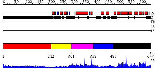
| Length: | 647 amino acids |
| Reference: | Drew K, et al. (2011) The proteome folding project: Proteome-scale prediction of structure and function. Genome Res. 2011 Sep 16 |
Listed below are up to the top 10 sequence alignment matches, by species, for the PSI-BLAST search against the protein sequence for FBpp0300193, RSG7-PA.
| Description | E-value | Query Range |
Subject Range |
|
|
600.0 | [0..196] | [639..5] |
|
Region A: Residues: [1-211] |
      1          11         21         31         41         51
      |          |          |          |          |          |
    1 MVTMNSEADK TQATNASSTA APAKTDCTSS NSSDPLAVAI PEPQSATAVV GGGAGGAAPG  60
   61 TATPSPTPLA NGSNNGTGSS VSPAATGTGA VATAASAPAA AGTTSASPPA SNPTTASTST 120
  121 ASVVGAKTAA ATTTSSSSAT SSSTAAAGSK QSGSSGGLLT VSGNHHHHHH AHPQQSAAGQ 180
  181 SAAQPPQPPT ATALAVPSAS HHQRGGKNED A
|
| Detection Method: |
Shown below is our most confident prediction for this domain.
Click here to view all matches.
Found no confident structure predictions for this domain.
|
Region A: Residues: [212-300] |
      1          11         21         31         41         51
      |          |          |          |          |          |
    1 PNILVYKKME AIIEKMQAES TGVAVRTVKA FMSKVPSVFT GADLVAWILK NFDVEDVTEA  60
   61 LHFAHLLSSH GYIFPIDDHA LTVKNDGTF
|
| Detection Method: | |
| Confidence: | 32.113509 |
| Match: | PF00610.12 |
| Description: | No description for PF00610.12 was found. |
Shown below is our most confident prediction for this domain.
Click here to view all matches.
Found no confident structure predictions for this domain.
|
Region A: Residues: [301-397] |
      1          11         21         31         41         51
      |          |          |          |          |          |
    1 YRFQTPYFWP SNCWEPENTD YAVYLCKRTM QNKTRLELAD YEAENLAKLQ KMFSRKWEFI  60
   61 FMQAESQSKV AKKRDKLERK VLDSQERAFW DVHRPMP
|
| Detection Method: |
Shown below is our most confident de novo (Rosetta) prediction for this domain.
Click here to view all matches.
Found no confident structure predictions for this domain.
|
Region A: Residues: [398-484] |
      1          11         21         31         41         51
      |          |          |          |          |          |
    1 GCVNTTEIDI KKAYRRGGSS HGTGSAGASL AKNPVEQLTR IIALRKQKLE RRTIKVSKAA  60
   61 EALVAYYDQY NEFDYFITSP ELPNPWQ
|
| Detection Method: | |
| Confidence: | 1.53 |
| Match: | 1xhmB |
| Description: | The Crystal Structure of a Biologically Active Peptide (SIGK) Bound to a G Protein Beta:Gamma Heterodimer |
Matching Structure (courtesy of the PDB): |
|
| Term | Confidence | Notes |
| binding | 1.90500749049821 | bayes_pls_golite062009 |
| signal transducer activity | 1.70315911322844 | bayes_pls_golite062009 |
| molecular transducer activity | 1.70315911322844 | bayes_pls_golite062009 |
| GTPase activity | 0.944482131496004 | bayes_pls_golite062009 |
| protein binding | 0.40736300061233 | bayes_pls_golite062009 |
| receptor binding | 0.363468238376214 | bayes_pls_golite062009 |
|
Region A: Residues: [485-647] |
      1          11         21         31         41         51
      |          |          |          |          |          |
    1 TDSTEMWDTE KNSKEVPVRR VKRWAFSLRE LLNDAIGREQ FTKFLEKEYS GENLKFWESV  60
   61 QEMKALPQSE IKEAIQKIWQ EFLAPEAPCP VNVDSKSVEL AREAVNSPSG PNRWCFDVAA 120
  121 SHVYHLMKSD SYSRYLRSDM YKDYLNCSRK KIKSIPNLFG VKR
|
| Detection Method: | |
| Confidence: | 56.154902 |
| Match: | 2es0A |
| Description: | Structure of the regulator of G-protein signaling domain of RGS6 |
Matching Structure (courtesy of the PDB): |
|