| Protein: | MSH7_ARATH |
| Organism: | Arabidopsis thaliana |
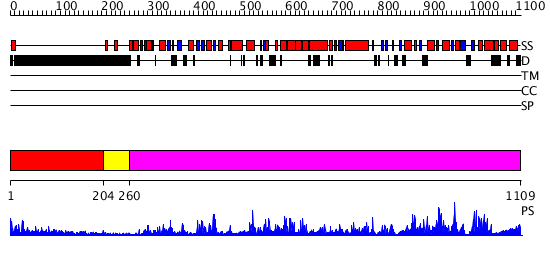
| Length: | 1109 amino acids |
| Reference: | Drew K, et al. (2011) The proteome folding project: Proteome-scale prediction of structure and function. Genome Res. 2011 Sep 16 |
No multiple sequence alignment data found for MSH7_ARATH.
|
Region A: Residues: [1-203] |
      1          11         21         31         41         51
      |          |          |          |          |          |
    1 MQRQRSILSF FQKPTAATTK GLVSGDAASG GGGSGGPRFN VKEGDAKGDA SVRFAVSKSV  60
   61 DEVRGTDTPP EKVPRRVLPS GFKPAESAGD ASSLFSNIMH KFVKVDDRDC SGERSREDVV 120
  121 PLNDSSLCMK ANDVIPQFRS NNGKTQERNH AFSFSGRAEL RSVEDIGVDG DVPGPETPGM 180
  181 RPRASRLKRV LEDEMTFKED KVP
|
| Detection Method: |
Shown below is our most confident prediction for this domain.
Click here to view all matches.
Found no confident structure predictions for this domain.
|
Region A: Residues: [204-259] |
      1          11         21         31         41         51
      |          |          |          |          |          |
    1 VLDSNKRLKM LQDPVCGEKK EVNEGTKFEW LESSRIRDAN RRRPDDPLYD RKTLHI
|
| Detection Method: |
Shown below is our most confident de novo (Rosetta) prediction for this domain.
Click here to view all matches.
Found no confident structure predictions for this domain.
|
Region A: Residues: [260-1109] |
      1          11         21         31         41         51
      |          |          |          |          |          |
    1 PPDVFKKMSA SQKQYWSVKS EYMDIVLFFK VGKFYELYEL DAELGHKELD WKMTMSGVGK  60
   61 CRQVGISESG IDEAVQKLLA RGYKVGRIEQ LETSDQAKAR GANTIIPRKL VQVLTPSTAS 120
  121 EGNIGPDAVH LLAIKEIKME LQKCSTVYGF AFVDCAALRF WVGSISDDAS CAALGALLMQ 180
  181 VSPKEVLYDS KGLSREAQKA LRKYTLTGST AVQLAPVPQV MGDTDAAGVR NIIESNGYFK 240
  241 GSSESWNCAV DGLNECDVAL SALGELINHL SRLKLEDVLK HGDIFPYQVY RGCLRIDGQT 300
  301 MVNLEIFNNS CDGGPSGTLY KYLDNCVSPT GKRLLRNWIC HPLKDVESIN KRLDVVEEFT 360
  361 ANSESMQITG QYLHKLPDLE RLLGRIKSSV RSSASVLPAL LGKKVLKQRV KAFGQIVKGF 420
  421 RSGIDLLLAL QKESNMMSLL YKLCKLPILV GKSGLELFLS QFEAAIDSDF PNYQNQDVTD 480
  481 ENAETLTILI ELFIERATQW SEVIHTISCL DVLRSFAIAA SLSAGSMARP VIFPESEATD 540
  541 QNQKTKGPIL KIQGLWHPFA VAADGQLPVP NDILLGEARR SSGSIHPRSL LLTGPNMGGK 600
  601 STLLRATCLA VIFAQLGCYV PCESCEISLV DTIFTRLGAS DRIMTGESTF LVECTETASV 660
  661 LQNATQDSLV ILDELGRGTS TFDGYAIAYS VFRHLVEKVQ CRMLFATHYH PLTKEFASHP 720
  721 RVTSKHMACA FKSRSDYQPR GCDQDLVFLY RLTEGACPES YGLQVALMAG IPNQVVETAS 780
  781 GAAQAMKRSI GENFKSSELR SEFSSLHEDW LKSLVGISRV AHNNAPIGED DYDTLFCLWH 840
  841 EIKSSYCVPK
|
| Detection Method: | |
| Confidence: | 1000.0 |
| Match: | 1wb9A |
| Description: | Crystal Structure of E. coli DNA Mismatch Repair enzyme MutS, E38T mutant, in complex with a G.T mismatch |
Matching Structure (courtesy of the PDB): |
|