| Protein: | zyg-12 |
| Organism: | Caenorhabditis elegans |
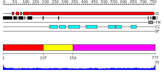
| Length: | 777 amino acids |
| Reference: | Drew K, et al. (2011) The proteome folding project: Proteome-scale prediction of structure and function. Genome Res. 2011 Sep 16 |
Listed below are up to the top 10 sequence alignment matches, by species, for the PSI-BLAST search against the protein sequence for zyg-12.
| Description | E-value | Query Range |
Subject Range |
|
|
272.0 | [0..5] | [777..1241] |
|
Region A: Residues: [1-206] |
      1          11         21         31         41         51
      |          |          |          |          |          |
    1 MLDLTNKESE SSDNGNSKYE DSIDGREVGT SKPFKEERSL EDLQADLADM AVWMEGLDAT  60
   61 KLPLNDPQLL CNGRAFSEVL HNVDKNFFTD GWLETMPENR TTNIMVFRSC TRKLWRKMFD 120
  121 YVNHINRTVV SSRWTDIHER IDGIYESDLP AMVNLGMAVV TLAHIGKNAK RFVDYSKALT 180
  181 STHKSMMSNV AKMVTTVIDE MPENPC
|
| Detection Method: | |
| Confidence: | 11.0 |
| Match: | 1wixA |
| Description: | The solution structure of RSGI RUH-026, conserved domain of HOOK1 protein from mouse |
Matching Structure (courtesy of the PDB): |
|
| Term | Confidence | Notes |
| binding | 1.77302826695282 | bayes_pls_golite062009 |
| protein binding | 1.34258839710246 | bayes_pls_golite062009 |
| microtubule binding | 0.26034159312323 | bayes_pls_golite062009 |
| tubulin binding | 0.11464228697646 | bayes_pls_golite062009 |
|
Region A: Residues: [207-358] |
      1          11         21         31         41         51
      |          |          |          |          |          |
    1 FHEISELHGS QSELNSLSES SGKLNGNGSS ERRSNADQIL VDAELEIERL RTETENQRKE  60
   61 IERLTKSFET AQHDMSSNSE SGDISILEKQ NEELRQKRRE LEEKNLELDA AVDQFKGIVF 120
  121 ELTNENDVLR RSDKERQRLQ TVLDAAQSDL DE
|
| Detection Method: | |
| Confidence: | 1.91 |
| Match: | 1jchA |
| Description: | Ribonuclease domain of colicin E3; Colicin E3 translocation domain; Colicin E3 receptor domain |
Matching Structure (courtesy of the PDB): |
|
|
Region A: Residues: [359-777] |
      1          11         21         31         41         51
      |          |          |          |          |          |
    1 WKTVANQYQK EAELSKQQDK EIKELLSQNK ALKSRLDHHV KSATLEDANK NGIAQLRTQV  60
   61 GGLTALNTEL KASLDSKKRC VEQLEIQLIQ HKEKVKELED RKDELIEERN RLENQLIFKE 120
  121 AVTPRSLHES MFEAGNLSFE PFSEKNTLPL EIENKRLTER IQELESLEPL KGELITLKSK 180
  181 NGVLEEEKLF ATKQIEELQQ QIEDLQENLL KNQEHASGDV VGLKIQLEKA EVEAQQMREA 240
  241 KMRAETNQAQ VDEILKKRTA ELEVNATALQ KAKAVIDELE YNSRPVSEDS MTSVQAFKEM 300
  301 KEENEKLRQK VEKLEIELNT VTQGFEQENR LLTSASHQQV LNRSIDEVMS MRAHAGSEEP 360
  361 QTLLDTQKMS GALPWRSLAS ETRRELPTAM ASILVLGFLV FIAWMFININ SALNAPPNA
|
| Detection Method: | |
| Confidence: | 17.0 |
| Match: | 1i84S |
| Description: | Heavy meromyosin subfragment |
Matching Structure (courtesy of the PDB): |
|