| Protein: | rpl-7A |
| Organism: | Caenorhabditis elegans |
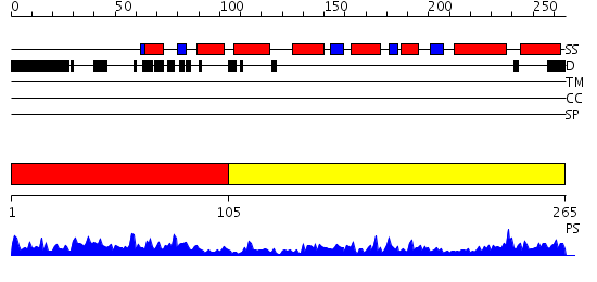
| Length: | 265 amino acids |
| Reference: | Drew K, et al. (2011) The proteome folding project: Proteome-scale prediction of structure and function. Genome Res. 2011 Sep 16 |
Listed below are up to the top 10 sequence alignment matches, by species, for the PSI-BLAST search against the protein sequence for rpl-7A.
| Description | E-value | Query Range |
Subject Range |
|
|
376.0 | [0..17] | [263..128] |
|
Region A: Residues: [1-104] |
      1          11         21         31         41         51
      |          |          |          |          |          |
    1 MPSKKVIKKK VAAVPAHIRA QTQVQKEVKN PLFEKRARNF NIGQDIQPKK DVTRFVKWPK  60
   61 YIRLQRQSAI LQKRLKVPPT INQFRTALDS QSARQAFKLL DKYR
|
| Detection Method: |
Shown below are all of our structure predictions for this domain.
Click here to view only most confident match.
| MCM Score |
GO Score |
GO Term |
SCOP Match |
SCOP Description | ||
| View | Download | 0.277 | N/A | N/A | a.40.1 | Calponin-homology domain, CH-domain |
| View | Download | 0.273 | N/A | N/A | a.138.1 | Multiheme cytochromes |
| View | Download | 0.233 | N/A | N/A | c.8.1 | Phosphohistidine domain |
| View | Download | 0.222 | N/A | N/A | d.112.1 | Phoshotransferase/anion transport protein |
|
Region A: Residues: [105-265] |
      1          11         21         31         41         51
      |          |          |          |          |          |
    1 PESTEAKKNR LRARAEARAA GKKEEVTKRP NTVRHGVNTI TRLVETRRAQ LVLIAHDVNP  60
   61 LEIVLHLPAL CRKYNVPYAI IKGKASLGTV VRRKTTAAVA LVDVNPEDKS ALNKLVETVN 120
  121 NNFSERHEEI RKHWGGGVMS AKSDAKKLKI ERARARDLGK L
|
| Detection Method: | |
| Confidence: | 20.0 |
| Match: | 1s1iG |
| Description: | Structure of the ribosomal 80S-eEF2-sordarin complex from yeast obtained by docking atomic models for RNA and protein components into a 11.7 A cryo-EM map. This file, 1S1I, Contains 60S subunit. The 40S Ribosomal Subunit Is In File 1S1H. |
Matching Structure (courtesy of the PDB): |
|