| Protein: | Dg-PA |
| Organism: | Drosophila melanogaster |
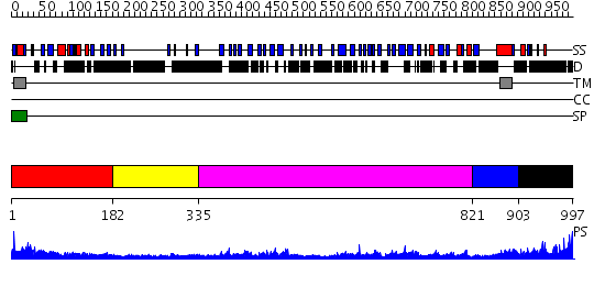
| Length: | 997 amino acids |
| Reference: | Drew K, et al. (2011) The proteome folding project: Proteome-scale prediction of structure and function. Genome Res. 2011 Sep 16 |
Listed below are up to the top 10 sequence alignment matches, by species, for the PSI-BLAST search against the protein sequence for Dg-PA.
| Description | E-value | Query Range |
Subject Range |
|
|
422.0 | [0..25] | [997..27] |
|
Region A: Residues: [1-181] |
      1          11         21         31         41         51
      |          |          |          |          |          |
    1 MRFQWFLSAS ASLSLFLLLD FVVWIHGERD FNVNDSQVPV IEPKDVPAYK QDPYVTELMS  60
   61 CQNTPSEIVL SLLLKKHDWS ELTATKRAHV QAKLAKFFAI PKEFISLDSV SKRELRSMHK 120
  121 LAMRKGGKGN KNIETLNRRL GRASFMIGCG PSYFVMGEPI AKQIAHQMKD GTIGALTEEN 180
  181 F
|
| Detection Method: | |
| Confidence: | 15.69897 |
| Match: | 1u2cA |
| Description: | Crystal Structure of a-dystroglycan |
Matching Structure (courtesy of the PDB): |
|
| Term | Confidence | Notes |
| binding | 1.57458227033664 | bayes_pls_golite062009 |
| cell adhesion molecule binding | 1.20941485091826 | bayes_pls_golite062009 |
| protein binding | 1.02797373424802 | bayes_pls_golite062009 |
| transporter activity | 0.851228093937992 | bayes_pls_golite062009 |
| transmembrane transporter activity | 0.677581850277121 | bayes_pls_golite062009 |
| receptor activity | 0.505108400296655 | bayes_pls_golite062009 |
| substrate-specific transporter activity | 0.483421965826537 | bayes_pls_golite062009 |
| signal transducer activity | 0.375469021130852 | bayes_pls_golite062009 |
| molecular transducer activity | 0.375469021130852 | bayes_pls_golite062009 |
| protein phosphatase binding | 0.13438543889553 | bayes_pls_golite062009 |
| phosphatase binding | 0.0613766967405507 | bayes_pls_golite062009 |
|
Region A: Residues: [182-334] |
      1          11         21         31         41         51
      |          |          |          |          |          |
    1 GLWFIWRKEL KSRSNRKRRQ SEGSGADEDD YDYGDDDEEV AEPSTEVPPV TTHAHRHHHG  60
   61 APNTVLNELE LSTQLPIDDF EGTATATQPT GTATATAIAS TAAAAAATAA AATATATTAN 120
  121 ASATTAGESE FHIESTTHRT ISSKEGGEPD AIS
|
| Detection Method: | |
| Confidence: | 1.08 |
| Match: | 1pk8A |
| Description: | Crystal Structure of Rat Synapsin I C Domain Complexed to Ca.ATP |
Matching Structure (courtesy of the PDB): |
|
|
Region A: Residues: [335-820] |
      1          11         21         31         41         51
      |          |          |          |          |          |
    1 SGNNSLANNE LSTPATPTSS VATTTASTPE SSSISSNFVS TDYMEPQPEE NSPPIIKTRL  60
   61 QKLAVTSGKA FTFHVLPETF YDAEDQGNLR LALTDKDGHE LKANSWLQFN ADKRELYGLP 120
  121 LDDTVSRWQY RLSATDSGNA SVTETVEISV QQHRAVRTIN HEISVFVRIN EKPGHNIDWQ 180
  181 LKLINAVART LDDSTNSAVV VREIRQTPHD PHSATFVYFN ETLPTSECPE KELKDIIARL 240
  241 DANRLSDLVQ PQLGIKSITG QLIGSCQKDL TQVKPTQHMT KNVPPMPRNQ VDRVNASLGQ 300
  301 LLVYKVPADT FYDANDNQLT LTLKTRDHLE LSPRHWLQFD SKNEEFYGIP KSGDIGSEEY 360
  361 LLVAEDSGGL SAHDALVVVV SPAPKRDFGF FFKAYLSIKH ERFNADLQRK FVERVAKLNG 420
  421 DPTTGQIQIR SITTHHDSDG TIVNFYNTTL YRKHNSCREK EVAMTRSVYL NSDLSLREAA 480
  481 KRALGP
|
| Detection Method: | |
| Confidence: | 31.0 |
| Match: | 1q55A |
| Description: | W-shaped trans interactions of cadherins model based on fitting C-cadherin (1L3W) to 3D map of desmosomes obtained by electron tomography |
Matching Structure (courtesy of the PDB): |
|
|
Region A: Residues: [821-902] |
      1          11         21         31         41         51
      |          |          |          |          |          |
    1 ELNLTNFSVV PFSICHHTEN IDTNQLDYIP SRPEEPTHKS SFGEDYMITF VWPIVIIVAM  60
   61 LVAASIIACC LHWCRQRSGK ME
|
| Detection Method: |
Shown below is our most confident de novo (Rosetta) prediction for this domain.
Click here to view all matches.
Found no confident structure predictions for this domain.
|
Region A: Residues: [903-997] |
      1          11         21         31         41         51
      |          |          |          |          |          |
    1 LGDEEERKSF RAKGIPVIFQ DEYEEKPEIG NKSPVILKDE KPPLLPPSYN TSNMNGDNDV  60
   61 DDYVPPPSVV VGGREVRGKS PATPSYRKPP PYVSP
|
| Detection Method: |
Shown below is our most confident prediction for this domain.
Click here to view all matches.
Found no confident structure predictions for this domain.