| Desc: |
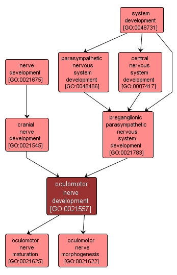
The process whose specific outcome is the progression of the oculomotor nerve over time, from its formation to the mature structure. This motor nerve innervates all extraocular muscles except the superior oblique and the lateral rectus muscles. The superior division supplies the levator palpebrae superioris and superior rectus muscles. The inferior division supplies the medial rectus, inferior rectus and inferior oblique muscles. This nerve also innervates the striated muscles of the eyelid. Pupillary constriction and lens movement are mediated by this nerve for near vision. In the orbit the inferior division sends branches that enter the ciliary ganglion where they form functional contacts (synapses) with the ganglion cells. The ganglion cells send nerve fibers into the back of the eye where they travel to ultimately innervate the ciliary muscle and the constrictor pupillae muscle. |