| Desc: |
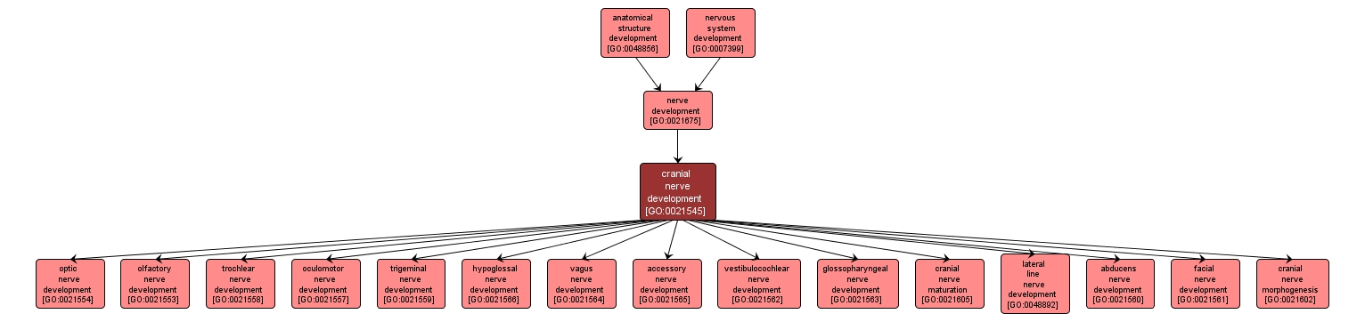
The process whose specific outcome is the progression of the cranial nerves over time, from its formation to the mature structure. The cranial nerves are composed of twelve pairs of nerves that emanate from the nervous tissue of the hindbrain. These nerves are sensory, motor, or mixed in nature, and provide the motor and general sensory innervation of the head, neck and viscera. They mediate vision, hearing, olfaction and taste and carry the parasympathetic innervation of the autonomic ganglia that control visceral functions. |