| Protein: | sym-2 |
| Organism: | Caenorhabditis elegans |
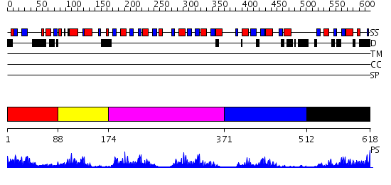
| Length: | 618 amino acids |
| Reference: | Drew K, et al. (2011) The proteome folding project: Proteome-scale prediction of structure and function. Genome Res. 2011 Sep 16 |
Listed below are up to the top 10 sequence alignment matches, by species, for the PSI-BLAST search against the protein sequence for sym-2.
| Description | E-value | Query Range |
Subject Range |
|
|
334.0 | [0..90] | [618..13] |
|
Region A: Residues: [1-87] |
      1          11         21         31         41         51
      |          |          |          |          |          |
    1 MNRQTSHPEQ RLLLIRVSDG NYDENTKLVV TSFYPRRQED ANNNSTSPSS ASSSGSEIST  60
   61 RLVNITPEDV VKFFDEHPTS SYIFTKG
|
| Detection Method: |
Shown below is our most confident de novo (Rosetta) prediction for this domain.
Click here to view all matches.
Found no confident structure predictions for this domain.
|
Region A: Residues: [88-173] |
      1          11         21         31         41         51
      |          |          |          |          |          |
    1 PEPIRHVLIP LFARNELCVP HPLHHYYDIK HLYYGNQEED GMEQEEIEDD VAIARAILDM  60
   61 DDELLEKTHQ FVNIEYQPAP ILEDQE
|
| Detection Method: | |
| Confidence: | 20.221849 |
| Match: | 1fjeB |
| Description: | Nucleolin |
Matching Structure (courtesy of the PDB): |
|
| Term | Confidence | Notes |
| binding | 2.30587477666777 | bayes_pls_golite062009 |
| nucleic acid binding | 1.2660519582186 | bayes_pls_golite062009 |
| protein binding | 0.88951142240317 | bayes_pls_golite062009 |
| RNA binding | 0.498012521772139 | bayes_pls_golite062009 |
|
Region A: Residues: [174-370] |
      1          11         21         31         41         51
      |          |          |          |          |          |
    1 VGADGDNVVC RARGLPWQAS DHHVAQFFAG LDIVPGGIAL CLSSEGRRNG EVLVQFSSQE  60
   61 SRDLALKRHR NFLLSRYIEV YKAGLDEFMH VATGSSTEAM EFVSANAIIV RMRGLPYDCT 120
  121 DAQIRTFFEP LKLTDKILFI TRTDGRPTGD AFVQFETEED AQQGLLKHRQ VIGQRYIELF 180
  181 KSTAAEVQQV VKRCNLI
|
| Detection Method: | |
| Confidence: | 45.30103 |
| Match: | 1pgzA |
| Description: | Crystal Structure of UP1 Complexed With d(TTAGGGTTAG(6-MI)G); A Human Telomeric Repeat Containing 6-methyl-8-(2-deoxy-beta-ribofuranosyl)isoxanthopteridine (6-MI) |
Matching Structure (courtesy of the PDB): |
|
| Term | Confidence | Notes |
| binding | 2.30587477666777 | bayes_pls_golite062009 |
| nucleic acid binding | 1.2660519582186 | bayes_pls_golite062009 |
| protein binding | 0.88951142240317 | bayes_pls_golite062009 |
| RNA binding | 0.498012521772139 | bayes_pls_golite062009 |
|
Region A: Residues: [371-511] |
      1          11         21         31         41         51
      |          |          |          |          |          |
    1 NSSPAVANAV EAPEEKKKDC VRLRGLPYEA TVQHIVTFLG DFATMVKFQG VHMVYNNQGH  60
   61 PSGEAFIQMI NEQAASACAA GVHNNFMSVG KKKRYIEVFQ ASAEELNLHH LVGQQHQVPP 120
  121 PAPLGFMGQL PPQAPQPQQF W
|
| Detection Method: | |
| Confidence: | 4.221849 |
| Match: | 2adbA |
| Description: | Solution structure of Polypyrimidine Tract Binding protein RBD2 complexed with CUCUCU RNA |
Matching Structure (courtesy of the PDB): |
|
|
Region A: Residues: [512-618] |
      1          11         21         31         41         51
      |          |          |          |          |          |
    1 SSYPSPPISP IVPGQVTQLI IYGIHMSIGV PELVANFTTP EHTVDNVLFT RWPTHLCPGE  60
   61 AILTLRNRGA PPPQTSPLSQ ISQLSAPNFA AYSHHPQNFP LQPILME
|
| Detection Method: | |
| Confidence: | 18.0 |
| Match: | 1u6fA |
| Description: | NMR solution structure of TcUBP1, a single RBD-unit from Trypanosoma cruzi |
Matching Structure (courtesy of the PDB): |
|
| Term | Confidence | Notes |
| binding | 2.30587477666777 | bayes_pls_golite062009 |
| nucleic acid binding | 1.2660519582186 | bayes_pls_golite062009 |
| protein binding | 0.88951142240317 | bayes_pls_golite062009 |
| RNA binding | 0.498012521772139 | bayes_pls_golite062009 |